|
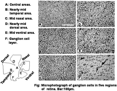
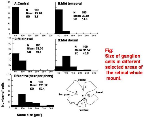
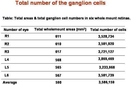
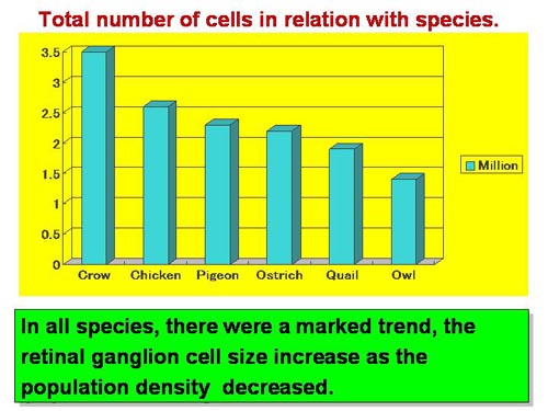
Distribution and Morphology of Retinal Ganglion cells of Jungle crow (Corvus macrorhynchos). ○Md.Lutfur Rahman1,2)・Masato Aoyama1)・Shoei Sugita1) (1Lab.of Function& Morphology,Dept. of Animal Science,Faculty of Agriculture, Utsunomiya University., 2Doctoral Course in United Graduate School of Agricultural Science, Tokyo University of Agriculture & Technology.) 【Abstract】 A retinal ganglion cell density map was generated using Nissl-stained retinal wholemounts from the jungle crow (Corvus macrorhynchos). The total number, distribution, and size of these cells were determined in the area centralis, and in temporal, nasal, dorsal, and ventral retinal regions. The mean total number of ganglion cells in the jungle crow retina was estimated to be 3.6 x 10⁶, with the highest density found in the area centralis (25,600/mm2) and dorso-temporal part of the retina, suggested the highest quality of vision, the density diminished nearly concentrically from the central area toward the retinal periphery. The number of ganglion cells was highest in the temporal retina, followed by the nasal, dorsal, and ventral retinal regions, respectively. Based on ganglion cell size, the retina seemed to consist of the following 5 regions: mid-central, nearly mid-temporal, mid-nasal, nearly mid-dorsal, and mid-ventral. Ganglion cell size ranged from 16-288 μm2, with smaller cells predominating in central regions above the optic disc and larger cells comprising more of the peripheral regions. The present study showed two areas of highly populated ganglion cells in crow’s retina, it is expected that, crow’s retina provides well developed monocular and binocular vision. Key words: area centralis, crow, ganglion cell, retina, soma size 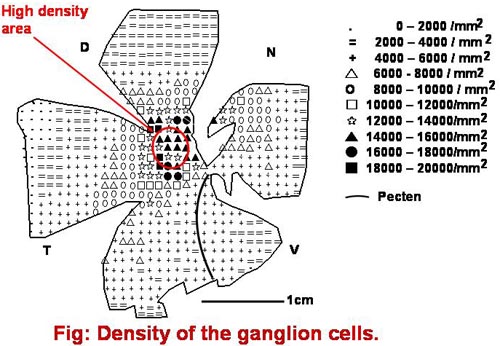
|